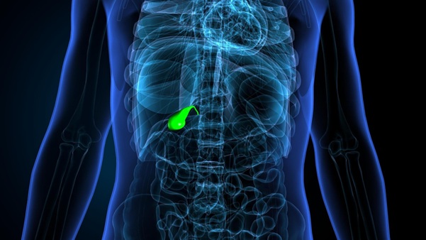
Gallbladder adenomyomatosis is a benign structural change that affects the wall of the gallbladder. In this condition, the inner lining of the gallbladder grows abnormally into the muscular layer beneath it, forming small pockets or outpouchings called Rokitansky-Aschoff sinuses. The gallbladder wall becomes thickened as a result.
The word sounds complicated, but the key takeaway is this: adenomyomatosis is not cancer. It is classified as a benign condition and is found fairly commonly during routine imaging.
That said, its presence does warrant proper evaluation. Depending on its pattern and any accompanying symptoms, your doctor will advise on whether monitoring or further management is appropriate.
Gallbladder adenomyomatosis and gallbladder thickening are related but not the same thing. Gallbladder thickening is a descriptive finding on imaging. It simply means the gallbladder wall appears thicker than normal, and there are many possible reasons for this, including inflammation, infection, heart failure, liver disease, or gallstones.
Gallbladder adenomyomatosis is one specific cause of gallbladder thickening. It has a distinct appearance on ultrasound or other imaging, often with characteristic features such as small cystic spaces or comet-tail artefacts that help distinguish it from other causes of wall thickening.
So while all cases of gallbladder adenomyomatosis involve some degree of wall thickening, not all gallbladder thickening is adenomyomatosis. This is why imaging interpretation by an experienced clinician is an important step in understanding what the finding actually means.
The exact cause is not fully understood. It is thought to develop over time as a result of increased pressure within the gallbladder, which may cause the inner lining to push into the muscular wall. Some researchers believe it may be related to chronic inflammation or changes that occur with age.
Gallbladder adenomyomatosis is more commonly found in:
1. Women than men
2. Adults over 40 years old
3. Individuals who also have gallstones
It is not considered hereditary, and there are no clear dietary or lifestyle factors definitively linked to its development.
Adenomyomatosis is categorised based on where the abnormal changes occur in the gallbladder.
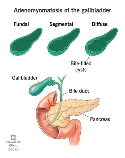
Image credit: Cleveland Clinic
Diffuse Type: Thickening affects the entire gallbladder wall and can sometimes be harder to distinguish from other conditions on imaging.
Fundal Type: The most common form, where thickening is limited to the tip of the gallbladder and is generally the easiest to identify and monitor.
Segmental Type: Thickening forms a ring around one section of the gallbladder, sometimes giving it an hourglass shape, and requires closer monitoring due to a low but noted association with gallbladder cancer in some studies.
Many people with gallbladder adenomyomatosis have no symptoms at all. It is frequently discovered incidentally during an ultrasound or scan done for another reason.
When symptoms do occur, or if you notice any of the following, consider consulting a doctor for further evaluation:
These symptoms are not unique to adenomyomatosis and can overlap with other gallbladder conditions such as gallstones or inflammation, which sometimes occur alongside it. This is why a proper assessment is important to understand what is actually causing the discomfort.
If you have already been told you have gallbladder adenomyomatosis and your symptoms are changing or worsening, it is worth seeing your doctor sooner rather than waiting for your next scheduled review.
Diagnosis is primarily made through imaging scans. The most common starting point is an abdominal ultrasound, which is painless, does not involve radiation, and is widely available. If the findings are unclear, further evaluation with MRI, CT imaging, or endoscopic ultrasound (EUS) may be considered to provide additional detail.
Once confirmed, treatment depends on your symptoms and scan findings.
If you have no significant symptoms, your doctor will likely recommend periodic monitoring with follow-up scans rather than immediate treatment.
If symptoms are persistent or the findings require closer attention, surgery to remove the gallbladder, known as a cholecystectomy, may be considered. In many cases, this is performed using a minimally invasive laparoscopic (keyhole) approach. Your surgeon will assess your condition and discuss the most appropriate treatment options.
The right approach is always decided on an individual basis based on your overall health, symptoms, and imaging results.
This article is intended for general educational purposes only and does not constitute medical advice. Please consult a qualified doctor for assessment and advice specific to your condition.
Copyright © ACE Specialist Surgery & Endoscopy | Terms & Conditions